Small ROI
Small Region of Interest (ROI) includes a series of data sets in which the sample is larger than the field of view (see table below). The related sample description and the experimental conditions are reported in tables below.
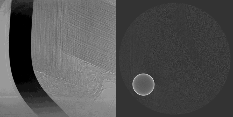
Double Bamboo Tin Wire
To load the data sets and perform a basic reconstruction using tomopy
tomopy recon --file-name tomo_00022.h5 --rotation-axis 980
tomopy recon --file-name tomo_00023.h5 --rotation-axis 893
tomopy recon --file-name tomo_00024.h5 --rotation-axis 972
tomo_ID |
00022 |
|---|---|
Image preview |
|
Download |
|
Instrument |
Elettra Syrmep |
Sample name |
Double Bamboo Tin Wire |
X-ray energy |
white beam mode |
Ring energy |
2 GeV |
Exposure time |
1 s |
Detector |
SCMOS 16-bit |
Sample-to-detector distance |
150 mm |
Pixel size |
2.4 µm |
Scan Range |
180 degree |
Number of Projections |
400 |
Rotation axis location |
980 |
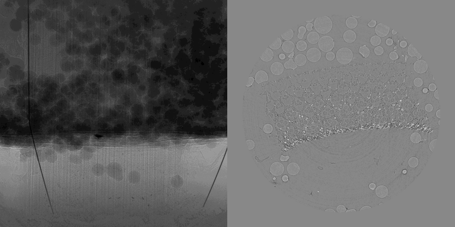
Bamboo Wires 2
tomo_ID |
00023 |
|---|---|
Image preview |
|
Download |
|
Instrument |
Elettra Syrmep |
Sample name |
Bamboo Wires 2 |
X-ray energy |
white beam mode |
Ring energy |
2 GeV |
Exposure time |
1 s |
Detector |
SCMOS 16-bit |
Sample-to-detector distance |
150 mm |
Pixel size |
2.6 µm |
Scan Range |
180 degree |
Number of Projections |
1800 |
Rotation axis location |
893 |
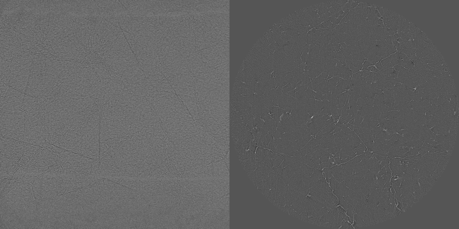
Polystyrene
tomo_ID |
00024 |
|---|---|
Image preview |
|
Download |
|
Instrument |
Elettra Syrmep |
Sample name |
Polystyrene |
X-ray energy |
white beam mode |
Ring energy |
2 GeV |
Exposure time |
1 s |
Detector |
SCMOS 16-bit |
Sample-to-detector distance |
150 mm |
Pixel size |
2.04 µm |
Scan Range |
180 degree |
Number of Projections |
400 |
Rotation axis location |
972 |
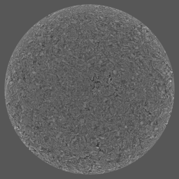
Electrode Particles
Detection and quantification of grey level differences of ca. 1% in the electrode particles – local tomography is making this challenging task even more difficult. Standard solutions used at the TOMCAT beamline (e.g. constant padding) are not sufficient in the phase retrieved case [B37], if small grey level differences need to be reliably detected.
To load the data sets and perform a basic reconstruction using tomopy
tomopy recon --file-name tomo_00070.h5 --rotation-axis 1289
To enable phase retrieval un-comment the appropriate setting in tomopy_rec.py
tomo_ID |
00070 |
|---|---|
Image preview |
|
Download |
|
Instrument |
SLS TOMCAT |
Sample name |
SLS_02 |
X-ray energy |
24.999 keV |
Sample-to-detector distance |
50 mm |
Scan Range |
180 degree |
Number of Projections |
1201 |
White Fields |
200 (100 before 100 after) |
Dark Fields |
10 |
Pixel size |
0.65 µm |
Rotation axis location |
1289.09525072 |
TomoLab
A second series of data sets (see table below) were acquired by microfocus CT at the TomoLab station of Elettra using a cone-beam geometry. The sample description and the experimental conditions are reported in tables below under tomo_00027 to tomo_00030.
Bone local
tomo_ID |
00027 |
|---|---|
Image preview |
|
Download |
|
Instrument |
TomoLab @Elettra |
Sample Name |
Bone local |
Voltage |
110 kV |
Current |
145 µA |
Filter |
0.1 mm Cu |
Focal spot |
16 µm |
Exposure time/proj |
4 s |
Detector |
CCD 12-bit |
Source-to-sample distance |
100 mm |
Source-to-detector distance |
250 mm |
Pixel size |
10.0 µm |
Number of Projections |
1800 |
Scan range |
360 degrees |
Start angle (degrees) |
0 |
Horizontal image size |
2004 pixels |
Vertical image size |
1335 pixels |
Detector horizontal pixel size |
25 µm |
Detector vertical pixel size |
25 µm |

Bone local stone
tomo_ID |
00028 |
|---|---|
Image preview |
|
Download |
|
Instrument |
TomoLab @Elettra |
Sample Name |
bone local stone |
Voltage |
60 kV |
Current |
250 µA |
Filter |
1.0 mm Al |
Focal spot |
16 µm |
Exposure time/proj |
4 s |
Detector |
CCD 12-bit |
Source-to-sample distance |
100 mm |
Source-to-detector distance |
250 mm |
Pixel size |
10.0 µm |
Number of Projections |
1800 |
Scan range |
360 degrees |
Start angle (degrees) |
0 |
Horizontal image size |
2004 pixels |
Vertical image size |
1335 pixels |
Detector horizontal pixel size |
25 µm |
Detector vertical pixel size |
25 µm |
Candy local
tomo_ID |
00029 |
|---|---|
Image preview |
|
Download |
|
Instrument |
TomoLab @Elettra |
Sample Name |
Candy local |
Voltage |
70 kV |
Current |
114 µA |
Filter |
1.5 mm Al |
Focal spot |
8 µm |
Exposure time/proj |
6 s |
Detector |
CCD 12-bit |
Source-to-sample distance |
100 mm |
Source-to-detector distance |
250 mm |
Pixel size |
10.0 µm |
Number of Projections |
1800 |
Scan range |
360 degrees |
Start angle (degrees) |
0 |
Horizontal image size |
2004 pixels |
Vertical image size |
1335 pixels |
Detector horizontal pixel size |
25 µm |
Detector vertical pixel size |
25 µm |
Smiling sample
tomo_ID |
00030 |
|---|---|
Image preview |
|
Download |
|
Instrument |
TomoLab @Elettra |
Sample Name |
Smiling sample |
Voltage |
60 kV |
Current |
300 µA |
Filter |
1.5 mm Al |
Focal spot |
20 µm |
Exposure time/proj |
1 s |
Detector |
CCD 12-bit |
Source-to-sample distance |
250 mm |
Source-to-detector distance |
350 mm |
Pixel size |
58.0 µm |
Number of Projections |
720 |
Scan range |
360 degrees |
Start angle (degrees) |
0 |
Horizontal image size |
668 pixels |
Vertical image size |
445 pixels |
Detector horizontal pixel size |
75 µm |
Detector vertical pixel size |
75 µm |